
![]()
Severe myopia
Severe myopia corresponds to above-normal growth of the eye, leading to elongation, strain and weakening of its various structures. It causes refractive visual problems that are corrected by glasses or contact lenses, but can also be the cause of specific pathologies.
Symptoms
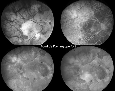
Myopic staphyloma
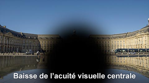
Strong myopes may experience areas of blindness known as scotomas. These are linked to atrophic areas of the retina in its posterior part, corresponding to the distension of the eyeball.
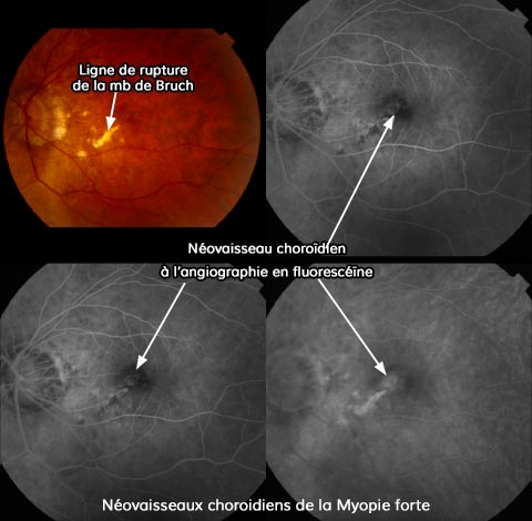
Choroidal neovessels
This distension can also lead to rupture of a membrane beneath the retina (Bruch’s membrane), resulting in communication with the vascularization of the choroid. In some cases, abnormal vessels (known as neovessels) develop beneath the retina, causing blood and fluid to accumulate. These vessels are different from those found in AMD. In this case, clinical symptoms include reduced central vision, distorted images and dark spots in the field of vision. A fundus examination combined with OCT will help make the diagnosis. Treatment involves intravitreal injections of anti-VEGF. One or two injections are usually sufficient to heal the neovessels, and the lesion does not chronically reactivate.
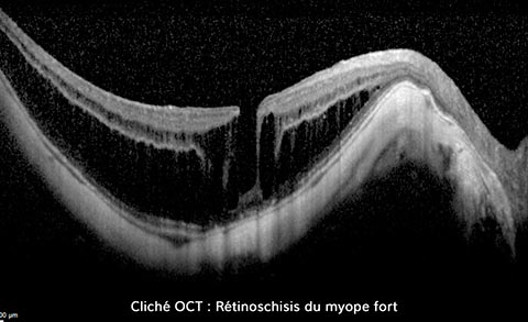
Epiretinal membranes and retinoschisis
Membranes on the surface of the retina occur more frequently in cases of severe myopia than in the general population. These membranes are often well tolerated, and do not require surgical removal as long as visual acuity is preserved. Some strong myopes may develop a specific anomaly of the central retina, known as retinoschisis. This anomaly corresponds to a “lamination” of the distended retina. It is discovered during an OCT examination of the retina. It does not require surgical treatment unless vision is severely impaired.
Retinal detachment
Retinal detachment is more frequent in strong myopes than in the general population, due to anatomical changes associated with the eye’s morphology and areas of fragility in the peripheral retina. Annual or biennial fundus monitoring is essential. This will enable the detection and preventive treatment (laser photocoagulation technique) of holes, tears or significant fragility that could develop into retinal detachment. If retinal detachment does occur, it is systematically treated surgically.