![]()
Ocular tumors
Uveal melanoma is a rare tumor: 500 to 600 new cases are diagnosed each year in France.
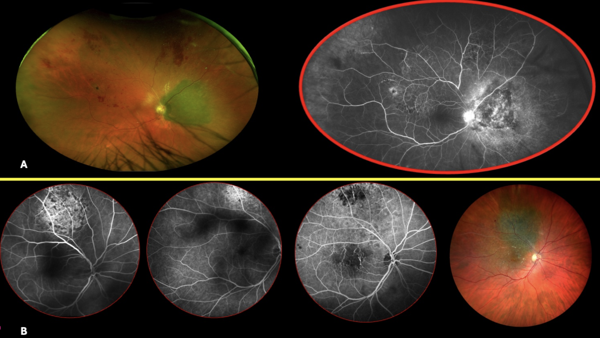
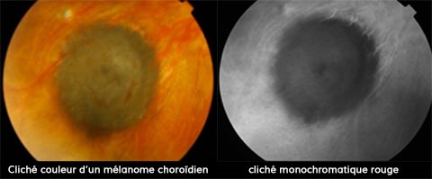
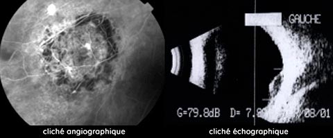
Melanoma is diagnosed by fundus examination.
Symptoms
The tumor may be completely asymptomatic, or may cause variable visual disturbances:
– flashes of light
– perception of floating bodies
– Amputation of the visual field
– Reduced visual acuity
Uveal melanomas appear as raised tumors, most often pigmented.
Various complementary examinations help confirm the diagnosis and guide the choice of treatment. These mainly involve angiography and ultrasound.
Evolution
After radiotherapy (proton therapy or radioactive plaque) of large tumors, complications may arise, in particular exudation or ocular hypertonia.
After excision surgery, possible complications are more precocious, notably intravitreal hemorrhage or retinal detachment.
Follow-up must be prolonged and rigorous, with ophthalmological and general examinations (liver ultrasound) every 3 and then 6 months.
The main problem lies in the overall evolution of the disease: despite a satisfactory ocular tumor response, metastatic disease is observed in a small percentage of cases. Chromosomal analysis of the tumour can be performed by genomic study, after transcleral needle puncture or transvitreal biopsy with a 27G TDC vitreotome.
The most common abnormalities found are monosomy 3 and multiplication of 8 in tumour cells.
There is a link between these anomalies and the patient’s vital prognosis.
The arrival on the market in 2022 of Tebentafusp (Kimmtrak, IMMUNOCORE), a bispecific antibody targeting HLA complexes, should double patients’ life expectancy.