
![]()
Central serous chorioretinopathy (CRSC)
Central Serous Chorioretinopathy (CSC) is a disease of the retina and choroid characterized by the presence of fluid under the retina. This fluid arises from a “leak point” in the pigment epithelium.
It is the fourth most common retinal disease.
It mainly affects young men (average age 45) (70% of cases), is favored by anxiety, and is aggravated or triggered by corticosteroids. General or local corticosteroid therapy should therefore be avoided whenever possible in these patients.
In women, it is aggravated by pregnancy.
Symptoms
When the macula is affected, patients experience a drop in visual acuity, the presence of a more or less dark spot in the central visual field (scotoma), image distortions (metamorphopsia) and a change in the size and color of objects.
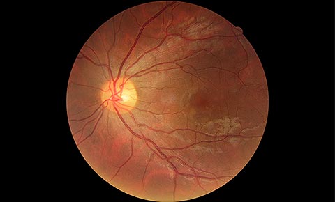
Fundus photography with CRSIt is performed during the patient’s clinical examination, which reveals a characteristic central fluid bubble in the fundus.
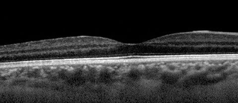
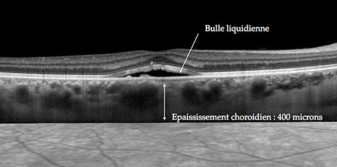
Choroidal thickeningThe OCT examination highlights the retinal serous
detachment bubble and, above all, makes it possible to follow its
evolution over time, as well as to measure the thickness of the choroidea,
the vascular layer beneath the retina. An increase in choroidal thickness is frequently observed, and this is one of the factors favoring the development of retinal detachment.
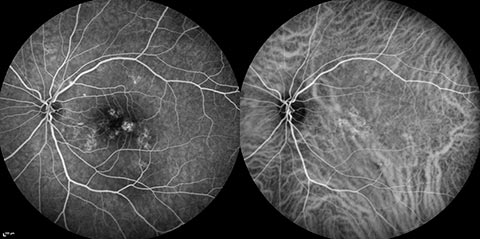
Fluorescence angiography and/or indocyanine green angiography may be indicated to confirm the diagnosis in case of doubt, rule out another disease and guide focal laser treatment when envisaged, by pinpointing the leakage point(s).
Diagnosis
Fundus photography with CRSIt is performed during the patient’s clinical
examination, which reveals a characteristic central fluid bubble in the
fundus.
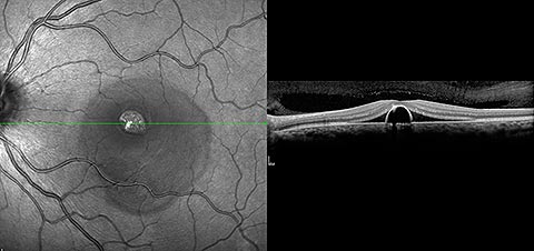
Choroidal thickeningThe OCT examination highlights the retinal serous
detachment bubble and, above all, makes it possible to follow its
evolution over time, as well as to measure the thickness of the choroidea,
the vascular layer beneath the retina. An increase in choroidal thickness is frequently observed, and this is one of the factors favoring the development of retinal detachment.
Fluorescence angiography and/or indocyanine green angiography may be indicated to confirm the diagnosis in case of doubt, rule out another disease and guide focal laser treatment when envisaged, by pinpointing the leakage point(s).
Treatment
It is considered in the absence of spontaneous healing after 4 months.
– Photocoagulation of the angiographically visible vanishing point(s) if these are outside the visual axis.
– Dynamic phototherapy with visudyne off-label.
Evolution
In most cases, the disease heals spontaneously within 1 to 4 months. This is the most common acute form in young people. It does not require treatment.
If there is no improvement after this period, treatment may be suggested. Sometimes treatment is offered earlier if the patient’s visual needs require it (for professional reasons, for example).
The SCCR may reoffend in the following months or years.
Some forms of CRSC have a chronic, recurrent course, affecting both eyes. They then constitute a pathology known as “diffuse retinal epitheliopathy”, with a reserved functional prognosis.